Delving into the realm of high power objective lens microscopy, this introduction immerses readers in a unique and compelling narrative that unravels the intricate workings of these powerful scientific tools. From their indispensable role in scientific research to their profound impact on medical diagnosis, high power objective lens microscopes have revolutionized our understanding of the microscopic world, opening up new avenues for exploration and discovery.
Composed of sophisticated optical components, including the objective lens, condenser, and eyepiece, these microscopes harness the principles of optics to magnify specimens, revealing intricate details and structures that would otherwise remain hidden from the naked eye. Each component plays a crucial role in the formation of a magnified image, working in harmony to provide researchers and scientists with unparalleled clarity and precision.
Introduction

A high power objective lens microscope is a scientific instrument designed to provide a highly magnified view of microscopic specimens. It is a crucial tool in scientific research and medical diagnosis, enabling scientists and medical professionals to observe and study the intricate details of cells, tissues, and microorganisms.
Importance of High Power Objective Lens Microscope
The high magnification capabilities of high power objective lens microscopes make them essential for various applications, including:
- Scientific Research:High power objective lens microscopes allow researchers to observe and study the ultrastructure of cells, including their organelles and molecular components. This enables them to investigate cellular processes, understand disease mechanisms, and develop new therapies.
- Medical Diagnosis:In the medical field, high power objective lens microscopes are used for diagnosing a wide range of diseases. They enable pathologists to examine tissue samples and identify abnormalities, such as the presence of cancerous cells or infectious microorganisms, aiding in accurate diagnosis and appropriate treatment.
Components of a High Power Objective Lens Microscope
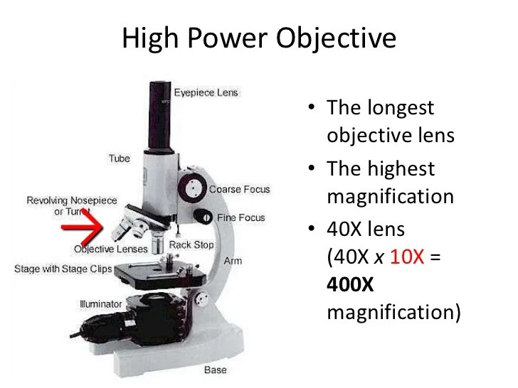
A high power objective lens microscope is a type of microscope that uses a high power objective lens to produce a magnified image of a specimen. The optical components of a high power objective lens microscope include the objective lens, condenser, and eyepiece.
Objective Lens
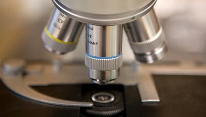
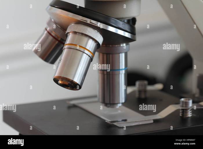
The objective lens is the most important optical component of a microscope. It is located at the bottom of the microscope and is responsible for gathering light from the specimen and focusing it on the eyepiece. The objective lens is typically made of glass or quartz and has a high numerical aperture (NA), which is a measure of its ability to gather light.
The higher the NA, the more light the objective lens can gather and the brighter the image will be.
Condenser
The condenser is located below the stage and is responsible for directing light from the light source onto the specimen. The condenser is typically made of glass or plastic and has a series of lenses that focus the light into a cone-shaped beam.
The cone of light is then directed onto the specimen, where it illuminates the specimen and allows the objective lens to gather light from it.
Eyepiece, High power objective lens microscope
The eyepiece is located at the top of the microscope and is responsible for magnifying the image produced by the objective lens. The eyepiece is typically made of glass or plastic and has a series of lenses that magnify the image.
The magnification of the eyepiece is typically measured in diopters (D), which is a measure of the power of the lens to magnify the image.
Types of High Power Objective Lens Microscopes

High power objective lens microscopes are available in various types, each offering unique advantages and disadvantages. These types include brightfield, darkfield, and phase contrast microscopes.
Brightfield Microscopes
Brightfield microscopes utilize transmitted light to illuminate the specimen. This creates a dark background against which the specimen appears bright. Brightfield microscopes are commonly used in routine laboratory applications and are suitable for observing stained specimens or those with inherent contrast.
- Advantages:Simple and inexpensive, easy to use, provides clear images of stained specimens.
- Disadvantages:Limited contrast for unstained specimens, can be difficult to visualize transparent or colorless specimens.
Darkfield Microscopes
Darkfield microscopes use oblique lighting to illuminate the specimen. This creates a dark background against which unstained specimens appear bright. Darkfield microscopes are particularly useful for observing live or unstained specimens, as they enhance the contrast of fine details.
- Advantages:High contrast for unstained specimens, ideal for visualizing transparent or colorless objects.
- Disadvantages:More complex and expensive than brightfield microscopes, can produce glare or halos around specimens.
Phase Contrast Microscopes
Phase contrast microscopes utilize a special condenser and objective lens to create phase shifts in the light passing through the specimen. This creates variations in brightness that correspond to differences in the specimen’s refractive index. Phase contrast microscopes are particularly useful for observing live cells and unstained specimens.
- Advantages:High contrast without the need for staining, ideal for visualizing transparent or colorless specimens, preserves the specimen’s natural state.
- Disadvantages:More complex and expensive than brightfield or darkfield microscopes, can produce halos or artifacts around specimens.
Applications of High Power Objective Lens Microscopes

High power objective lens microscopes are indispensable tools in various scientific fields, enabling researchers and scientists to make groundbreaking discoveries. Their ability to magnify specimens at exceptionally high resolutions allows for the visualization and analysis of minute details, providing valuable insights into the structure and function of biological systems, materials, and more.
In biology, high power objective lens microscopes are crucial for studying cellular and subcellular structures. They enable researchers to observe the morphology, dynamics, and interactions of organelles, proteins, and DNA molecules, providing insights into cellular processes and the development of diseases.
In medicine, high power objective lens microscopes are used for diagnostic purposes, such as identifying pathogens in tissue samples or detecting abnormalities in cells. They also play a vital role in surgical procedures, allowing surgeons to visualize fine structures and perform precise manipulations.
In materials science, high power objective lens microscopes are employed to characterize the microstructure of materials, including their grain size, crystal structure, and defects. This information is essential for understanding the mechanical properties, electrical conductivity, and other performance characteristics of materials, enabling the development of new and improved materials for various applications.
Maintenance and Troubleshooting

High power objective lens microscopes are delicate instruments that require proper maintenance to ensure optimal performance and longevity. Regular cleaning, alignment, and calibration are essential to prevent damage and ensure accurate results.
Common problems that can occur with high power objective lens microscopes include:
Cleaning Procedures
- Lens cleaning:Use lens paper or a soft, lint-free cloth to gently wipe away dust or debris from the lens surfaces. Avoid using abrasive materials or solvents.
- Body cleaning:Clean the microscope body with a damp cloth and mild detergent. Avoid using harsh chemicals or solvents.
- Stage cleaning:Clean the stage with a soft cloth and isopropyl alcohol to remove any contaminants.
Alignment and Calibration
- Köhler illumination alignment:Adjust the condenser and diaphragm to achieve even illumination of the specimen.
- Condenser centering:Center the condenser under the objective lens to optimize illumination.
- Objective lens calibration:Calibrate the objective lenses using a microscope calibration slide to ensure accurate magnification and resolution.
Troubleshooting
- Blurry images:Check for proper alignment of the microscope components, clean the lenses, and adjust the focus.
- Uneven illumination:Adjust the Köhler illumination settings or check for condenser alignment.
- Mechanical issues:Inspect the microscope for any loose or damaged parts, such as the stage or focusing mechanism.
- Electrical problems:Check the power supply and connections to ensure the microscope is receiving adequate power.
Advancements in High Power Objective Lens Microscopy: High Power Objective Lens Microscope
High power objective lens microscopy has seen significant advancements in recent years, pushing the boundaries of what is possible in the field of microscopy. These advancements have led to the development of new and improved microscopes, as well as new techniques that have opened up new possibilities for research.
One of the most significant advancements in high power objective lens microscopy is the development of super-resolution microscopes. These microscopes use advanced optical techniques to achieve resolutions that are far beyond the diffraction limit of conventional microscopes. This allows researchers to visualize and study biological structures with unprecedented detail, opening up new possibilities for research in fields such as cell biology and neuroscience.
Super-Resolution Microscopy Techniques
There are several different super-resolution microscopy techniques that have been developed, each with its own advantages and disadvantages. Some of the most common techniques include:
- STED microscopy: This technique uses a donut-shaped laser beam to excite a small region of the sample. The fluorescence from this region is then detected and used to create an image with a resolution that is much higher than the diffraction limit.
- PALM microscopy: This technique uses a series of short laser pulses to activate individual molecules within the sample. The fluorescence from these molecules is then detected and used to create an image with a resolution that is much higher than the diffraction limit.
- SIM microscopy: This technique uses a structured illumination pattern to excite the sample. The fluorescence from the sample is then detected and used to create an image with a resolution that is twice the diffraction limit.
These are just a few of the many advancements that have been made in high power objective lens microscopy in recent years. These advancements are continuing to push the boundaries of what is possible in the field of microscopy, and they are opening up new possibilities for research in a wide range of fields.
Question Bank
What are the advantages of using high power objective lens microscopes?
High power objective lens microscopes offer several advantages, including:
- Higher magnification, allowing for the visualization of smaller structures and details.
- Improved resolution, providing sharper and clearer images.
- Increased contrast, enhancing the visibility of specific features.
- Specialized techniques, such as phase contrast and darkfield microscopy, which reveal additional information about specimens.
What are the different types of high power objective lens microscopes?
There are several types of high power objective lens microscopes, each with its own advantages and applications:
- Brightfield microscopes: Provide clear and detailed images of stained or unstained specimens.
- Darkfield microscopes: Utilize oblique lighting to enhance the visibility of unstained specimens.
- Phase contrast microscopes: Reveal transparent or colorless specimens by converting phase differences into visible contrast.
- Super-resolution microscopes: Push the limits of resolution, enabling the visualization of structures below the diffraction limit.
What are the applications of high power objective lens microscopes?
High power objective lens microscopes find applications in a wide range of fields, including:
- Biology: Studying cellular structures, microorganisms, and tissue samples.
- Medicine: Diagnosing diseases, examining tissue biopsies, and guiding surgical procedures.
- Materials science: Characterizing materials, analyzing defects, and developing new materials.
- Forensic science: Examining evidence, identifying trace materials, and analyzing fingerprints.