You mistakenly confuse the primary stain and counterstain, a common pitfall in histological staining that can lead to misinterpretations and inaccurate tissue diagnosis. This guide will delve into the intricacies of primary and counterstain identification, empowering you with the knowledge to avoid confusion and ensure reliable histological analysis.
In this comprehensive exploration, we will unravel the typical mistakes associated with primary and counterstains, highlighting the consequences of misidentification and the significance of accurate stain identification. We will also provide practical tips and techniques for stain differentiation, troubleshooting common pitfalls, and best practices for reliable stain identification.
Primary Stain Confusion: You Mistakenly Confuse The Primary Stain And Counterstain

Primary stains, which impart color to specific tissue components, are often mistaken for counterstains. This confusion arises due to similarities in their staining characteristics, such as color or binding properties.
Common primary stains that are frequently misidentified as counterstains include:
- Hematoxylin (blue-purple nuclear stain) mistaken for methylene blue (counterstain)
- Eosin (pink cytoplasmic stain) mistaken for phloxine (counterstain)
Misidentifying primary and counterstains can lead to incorrect tissue interpretation and diagnostic errors.
Counterstain Confusion, You mistakenly confuse the primary stain and counterstain
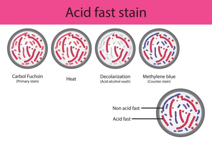
Counterstains provide contrasting colors to primary stains, enhancing tissue visibility and facilitating structural analysis. Common mistakes in identifying counterstains include:
- Mistaking a counterstain for a primary stain due to similar staining intensity or color.
- Assuming that all blue or red stains are counterstains, regardless of their binding properties.
Counterstains play a crucial role in histological staining by providing contrast and highlighting specific tissue components. Distinguishing them from primary stains is essential for accurate tissue interpretation.
Impact on Histological Interpretation
Mistaking primary and counterstains can significantly impact tissue diagnosis and lead to misinterpretations:
- Overestimating or underestimating the presence of certain tissue components.
- Misinterpreting the type of tissue or pathological process.
Accurate stain identification is crucial for reliable histological analysis and correct tissue diagnosis.
Best Practices for Stain Identification
To avoid confusion between primary and counterstains, consider the following key factors:
- Stain color: Primary stains typically impart specific colors to tissue components, while counterstains provide contrasting colors.
- Binding properties: Primary stains bind specifically to target molecules, while counterstains bind non-specifically to background structures.
Additional tips for distinguishing between primary and counterstains include:
- Review the staining protocol to determine the specific stains used.
- Consult reference materials or consult with an experienced histologist.
Troubleshooting Stain Confusion
If confusion arises, the following troubleshooting guide can help resolve the issue:
- Re-examine the staining protocol and ensure that the correct stains were used.
- Compare the staining results with known positive and negative controls.
- Consider using alternative staining methods or specific blocking agents to enhance stain differentiation.
By following these guidelines, histologists can avoid confusion between primary and counterstains, ensuring accurate tissue interpretation and reliable diagnostic outcomes.
Clarifying Questions
What are the most common mistakes in identifying primary and counterstains?
Mistaking a primary stain for a counterstain and vice versa is a common error, especially for beginners in histology.
How can I avoid confusion between primary and counterstains?
Pay attention to the color, binding properties, and intended use of the stains. Primary stains typically bind specifically to target molecules, while counterstains provide contrasting background.
What are the consequences of misidentifying primary and counterstains?
Misidentification can lead to misinterpretation of tissue structures, incorrect diagnosis, and unreliable histological analysis.

